Heart Defect PDA

In a normal, healthy heart, the two main blood vessels, namely the aorta, perfusing the body and the pulmonary artery, perfusing the lungs, are separated. It should be noted that the blood pressure in the aorta is much higher than the blood pressure in the pulmonary artery.

During pregnancy, the child’s lungs are not used for oxygenation, therefore the blood does not need to go through the lungs but is guided to the aorta from the pulmonary artery through a large vessel, called the ductus arteriosus. This changes immediately after birth when the lungs are filled with air. Normally the ductus arteriosus closes within the first hours of life.

A patent ductus arteriosus (PDA) is a situation where a neonate’s ductus arteriosus fails to close after birth and remains “patent”. This PDA is one of the more common congenital heart defects. A PDA is much more common among premature babies and infants with genetic disorders than others. In many children the PDA will sometimes shrink and go away without any help. Most defects close within two years without treatment being necessary. 

PDA

Because of the pressure difference between the two vessels there is a variable amount of blood flowing at a high pressure from the aorta to the pulmonary artery and therefore to the lungs. If too much blood flows into the lungs, this puts a strain on the heart and increases the blood pressure in the pulmonary arteries. If the defect is very large the lungs could become overloaded with blood and fluid, which can result in an enlarged heart and even heart failure. When there is significant blood flow to the lungs, the PDA needs to be closed. 

When a patient has a PDA, the following symptoms can occur:

A. If the PDA is small, there might not be any symptoms, therefore some patients will not be diagnosed until later in childhood. 

B. If the PDA is of moderate size, children may show nonspecific symptoms such as:
  • Tiredness and Sweating
C. If the PDA is big, children suffer from the blood volume overloading the heart and lungs. They can show:
  • Strong and forceful pulse
  • Rapid breathing
  • Poor feeding habits
  • Shortness of breath
  • Poor growth

Closure of the PDA

There are different ways to treat a PDA defect. At first, the symptoms from the PDA defect can be regulated with medication however this is only a transient method without treating the underlying cause. Other ways to treat a PDA defect are by heart surgery or catheter-based procedures, i.e. interventional treatment. 

Interventional closure

The closure is performed in a cardiac catheterization laboratory, known as a cath lab. Techniques vary between clinics and physicians active in catheter interventions. In older patients the procedure can be done under local anesthesia. In younger patients, patients who are afraid and, in all children, the procedure is performed with the support of sleeping medication (sedation) or by general anesthesia. The procedure itself will be performed by a specialist in treating congenital heart defects, a (pediatric) cardiologist.

A thin tube – a catheter – is inserted into a blood vessel in the groin and guided to the heart. As the shape of a PDA can vary substantially, the size, diameter and length of the PDA is measured exactly by angiography. Thereafter a special closure device, a PDA occluder is chosen. The procedure is constantly monitored by X-ray. The catheter is then exchanged for a larger catheter and a delivery sheath is placed across the PDA. The appropriate device, a PDA occluder, is pushed through the delivery sheath to be placed correctly inside the PDA to close the defect. After placement, the physician will check the position by repeat angiography, release the device and withdraw the delivery sheath. Typically the procedure takes around 60 minutes. The defect is now closed and the tissue will grow around the device. 

Occluders and airports

The device will not set-off any metal detectors at an airport security scan. 

If you have additional questions or need additional information, please contact Occlutech’s service center via email: info@occlutech.com

Airport

Illustrations of PDA

PDA area shown on a normal, healthy heart

In a normal, healthy heart, the two main blood vessels, namely the aorta, perfusing the body and the pulmonary artery, perfusing the lungs, are separated. It should be noted that the blood pressure in the aorta is much higher than the blood pressure in the pulmonary artery.

A heart with a patent ductus arteriosus (PDA)
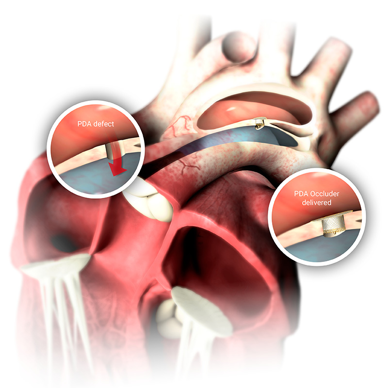
During pregnancy, the child’s lungs are not used for oxygenation, therefore the blood does not need to go through the lungs but is guided to the aorta from the pulmonary artery through a large vessel, called the ductus arteriosus.

This changes immediately after birth when the lungs are filled with air. Normally the ductus arteriosus closes within the first hours of life.

Patent ductus arteriosus (PDA) is a situation where a neonate’s ductus arteriosus fails to close after birth and remains “patent”. This PDA is one of the more common congenital heart defects. Because of the pressure difference between the two vessels there is a variable amount of blood flowing at a high pressure from the aorta to the pulmonary artery and therefore to the lungs. When there is significant blood flow to the lungs, the PDA needs to be closed.  

Closing the PDA defect
Usually the closure can be performed without surgery and is performed in a cardiac catheterization laboratory, known as a cath lab. A thin tube – a catheter – is inserted into a blood vessel in the groin and guided to the heart. As the shape of a PDA can vary substantially, the size, diameter and length of the PDA is measured exactly by angiography.

Thereafter, a special closure device, a PDA occluder is chosen. The catheter is then exchanged for a larger catheter and the delivery sheath is placed across the PDA. The PDA occluder is then pushed through the delivery sheath and placed correctly inside the PDA to close the defect. After placement the physician will check the position by repeat angiography, release the device and withdraw the delivery sheath. 

Occlutech PDA device in position
After exact placement the device is now in the correct position and the defect is closed. No more blood can flow cross the PDA and harm the lungs. After only a few months the occluder will be covered by a thin layer of blood vessel cells, then covered by the tissue of the heart and over time the defect will be completely overgrown. Only an X-ray and ultrasound (ECHO) will show that a procedure has been carried out. 

PDA Occluder

The Occlutech PDA Occluder has been developed specifically for the minimally invasive, transcatheter closure of Patent Ductus Arteriosus (PDA). PDA is a congenital abnormality of the heart in which blood is shunted from the aorta (high pressure vessel) through the ductus arteriosus into the pulmonary artery (lower pressure vessel) resulting in fluid build-up in the lungs.

The device is made of braided Nitinol threads. Nitinol is a metal alloy of nickel and titanium with unique features, including superelasticity and shape memory properties. Each Occlutech PDA Occluder consists of an aortic disk connected to a conical-shaped shank. To accommodate different PDA anatomies, the Occlutech PDA Occluder is available in several sizes, each with either a standard (short), or long shank. Polyethylene (PET) patches inside the device support immediate closure of the defect. 

occlutech PDA